You’ve probably felt it at some point. The afternoon energy crash that hits regardless of what you ate. The training plateau where effort no longer yields the results it once did. The blood sugar that swings more than it should, or the feeling that your body burns carbohydrates fine but can’t seem to sustain fat as a fuel for long.
These are the felt symptoms of something the research literature calls metabolic inflexibility — the impaired ability to switch between fuel sources depending on what’s available. And inside your cells, one family of molecules has emerged as one of the most informative windows into whether that switching system is working.
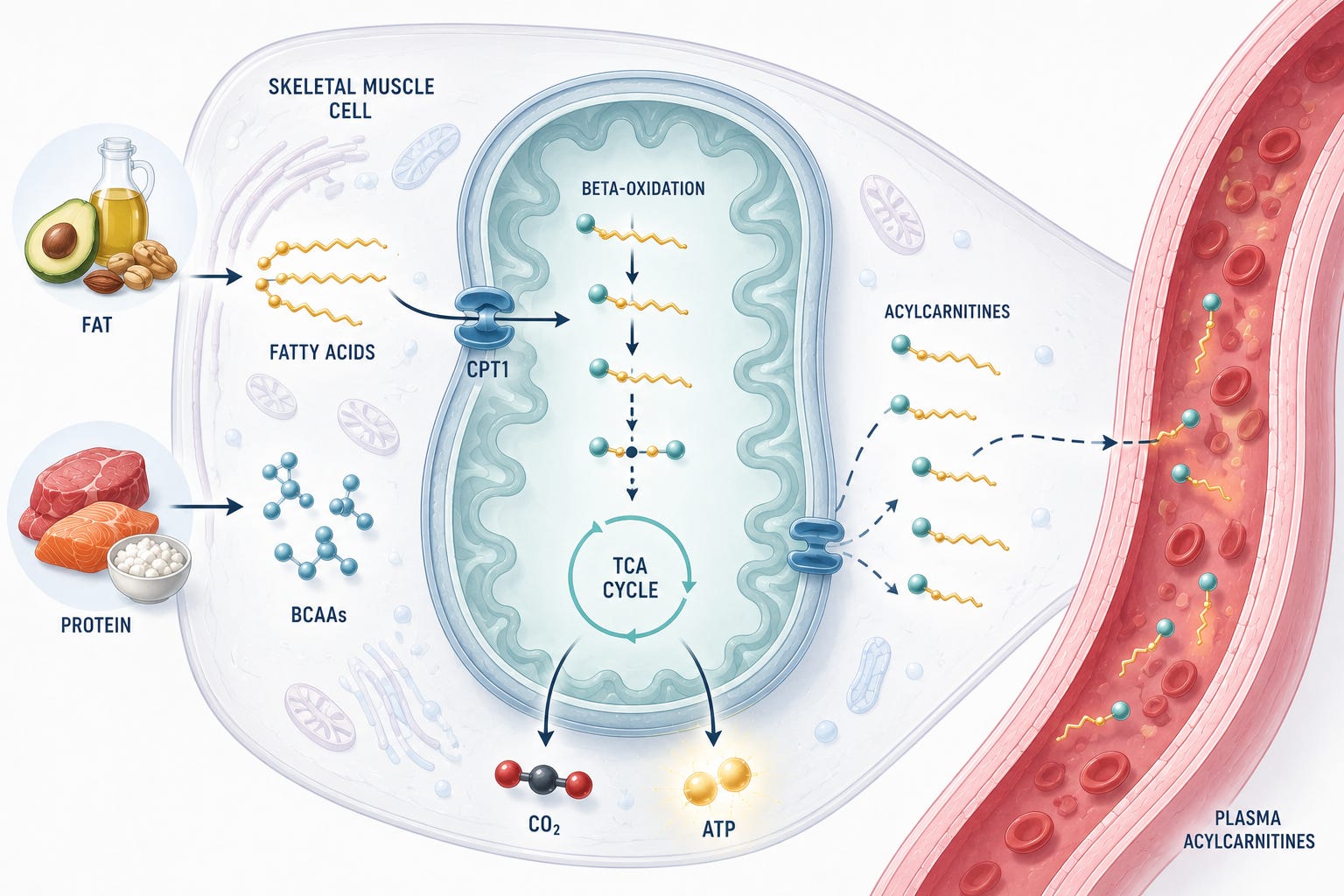
They are called acylcarnitines. Most people have never heard of them. They don’t appear on standard metabolic panels. But they have been the subject of an expanding body of research identifying them as consistent markers of metabolic dysfunction — and understanding what they are and what they reflect changes how you think about metabolic health monitoring entirely.
The fuel-switching problem
Your body runs on two primary fuels: glucose and fatty acids. Which one dominates at any moment depends on the situation — whether you’ve just eaten, how hard you’re exercising, how much glycogen is available, what hormonal signals are circulating. A metabolically flexible system shifts smoothly between these fuels, burning mostly carbohydrates after a meal and shifting toward fat during fasting or sustained low-intensity effort.
Metabolic inflexibility is what happens when that switching mechanism breaks down. In people with obesity and type 2 diabetes, the transition from fat oxidation to carbohydrate oxidation is measurably blunted, though evidence suggests this may be primarily a consequence of impaired glucose transport rather than a primary oxidative defect. ¹ The downstream consequences extend beyond energy management: impaired fuel switching is tightly linked to insulin resistance, the condition in which cells stop responding normally to insulin’s signal to take up glucose.
This is where acylcarnitines enter the story — not as the problem itself, but as a readout of a system under strain.
What acylcarnitines are — and where they come from
Long-chain fatty acids cannot cross the inner mitochondrial membrane on their own. To enter the mitochondria — where fat oxidation actually occurs — they must first be conjugated to a molecule called carnitine. The enzyme that drives this reaction, carnitine palmitoyltransferase 1 (CPT1), sits at the outer mitochondrial membrane and acts as the gatekeeper: it controls the rate at which fatty acids enter the organelle for burning.²
Once inside, fatty acids undergo beta-oxidation — a sequential process that strips two carbons at a time, generating acetyl-CoA, which then feeds into the tricarboxylic acid (TCA) cycle to produce energy. The acylcarnitine intermediates produced along this pathway are normally short-lived. When the system runs efficiently, they are fully processed, and the carnitine is recycled.
The problem arises when the input exceeds the throughput. In states of chronic fat overload — sustained high-fat dietary exposure, obesity, or conditions that impair mitochondrial capacity — fatty acid flux into mitochondria outpaces the TCA cycle’s ability to process acetyl-CoA. The result is a bottleneck: incomplete beta-oxidation, with acylcarnitine intermediates accumulating rather than being fully metabolized.² These accumulated intermediates are exported from the cell and appear in the blood. That is what you measure in a plasma acylcarnitine profile — the exhaust of a metabolic signature that is working hard but not working completely through the energy-producing machinery.³
The branched-chain amino acids (BCAA) connection adds another layer. BCAA — leucine, isoleucine, and valine — are catabolized through a separate pathway that converges on the same mitochondrial machinery. Their breakdown generates short-chain acylcarnitines, particularly C3 (propionylcarnitine) and C5 (isovalerylcarnitine). When BCAA catabolism is impaired, as it is in insulin-resistant states, these species accumulate alongside the products of incomplete fatty acid oxidation.³ The acylcarnitine profile, in this sense, is capturing two dysfunctional systems at once — and their signals are partially intertwined.
Diet shapes the input to this system directly. A chronically high-fat, calorie-dense dietary pattern floods the mitochondria with substrate. Adiposity adds circulating free fatty acids to this load. Genetics influence mitochondrial capacity and the enzymes involved in beta-oxidation and BCAA catabolism. And prior exercise history shapes the mitochondrial infrastructure available to handle the load.²
What the measurement is actually capturing
Acylcarnitines are measured by liquid chromatography-tandem mass spectrometry (LC-MS/MS), the same platform used for ceramide profiling. The analysis distinguishes species by chain length and the nature of the acyl group — short-chain (C2–C6), medium-chain (C8–C14), and long-chain (C16+) acylcarnitines have different biological origins and different clinical associations.³
This is where the measurement context matters enormously — and where a single number without context tells an incomplete story.
Acylcarnitines are acutely and transiently responsive to physiological state. During exercise, plasma acylcarnitines rise substantially — the liver releases short-chain species, the exercising muscle releases medium-chain species, and both return to baseline during recovery.⁴ This is normal, expected physiology. A measurement taken two hours post-exercise reflects a very different biological state than a measurement taken after an overnight fast, even in the same person. The fasting state itself elevates long-chain acylcarnitines—this too is a normal metabolic response, not a disease signal. During fasting, the body naturally switches to a mode of higher fatty acid oxidation to meet energy demands, which leads to a physiological rise in circulating acylcarnitine esters.4,5 This reflects the system’s inherent flexibility to ramp up fat-burning machinery when glucose is unavailable. ⁴
What differentiates a chronic disease signal from this acute response is the lack of resolution. In healthy metabolism, these levels are transient and return to baseline upon refeeding.3 In metabolically inflexible individuals, the system remains ‘stuck’; long-chain acylcarnitines may be chronically elevated even in the fasted state, often appearing alongside a bottleneck of short- and medium-chain species that indicates the mitochondrial engine is failing to process the fat load completely.3
It is also worth noting that the liver and skeletal muscle contribute differently to what appears in plasma. The liver is the primary source of circulating short-chain acylcarnitines in the fasted state; muscle releases predominantly medium-chain species during exercise.⁴ Plasma measurements integrate signals from multiple tissues simultaneously — which is both their strength (a whole-body readout) and a limit on interpretation (you cannot tell from plasma alone which tissue is the primary contributor).
What the evidence actually shows
The observation that something was distinctly different about the acylcarnitine profiles of people with insulin resistance came into sharp focus in a 2009 study published in Cell Metabolism. Using targeted metabolomics in skeletal muscle and plasma from obese and lean subjects, Koves and colleagues found that insulin resistance in skeletal muscle was not, as previously assumed, caused by insufficient fat oxidation. Instead, it was characterized by excessive beta-oxidation that outpaced TCA cycle capacity — generating accumulated acylcarnitine intermediates that correlated with impaired insulin signaling.⁵ This was the foundational reframe: acylcarnitines were not a marker of fat underutilization, but of fat overutilization that the cell couldn’t complete through the energy-producing machinery.
The same year, Newgard and colleagues published a landmark metabolomics study in Cell Metabolism linking a BCAA-related metabolite signature — including branched-chain acylcarnitines — to obesity and insulin resistance in humans. Critically, this study demonstrated a mechanism: in rats, adding BCAA supplementation to a high-fat diet produced insulin resistance accompanied by accumulation of multiple acylcarnitines in muscle, mediated through mTOR and JNK signaling.⁶ The human metabolite signature wasn’t merely descriptive — it tracked a biologically active process.
The population-level evidence reinforced this. A meta-analysis published in Diabetes Care pooled data from 61 prospective metabolomics studies involving 71,196 participants and 11,771 incident type 2 diabetes cases. Specific acylcarnitines — including C4-OH, C5, C5-OH, and C8:1 — were significantly associated with type 2 diabetes risk across cohorts, with hazard ratios ranging from 1.07 to 2.58 per standard deviation.⁷ This is not one study finding an association. It is the consistent signal across dozens of independent cohorts.
Now the honest comparison, which this series commits to making. Branched-chain amino acids have the strongest metabolomics-derived predictive signal for type 2 diabetes in this metabolite class — stronger than acylcarnitines alone. Wang and colleagues, using the Framingham Offspring cohort, followed 2,422 normoglycemic individuals for 12 years and found that a combination of five amino acids including three BCAAs predicted diabetes with more than fivefold higher risk in the top quartile — replicated in an independent cohort.⁸ A subsequent Mendelian randomization analysis of 47,877 cases and more than 267,000 controls established that genetically predicted BCAA elevation is strongly consistent with a causal role in type 2 diabetes risk, according to Mendelian randomization studies, with odds ratios of 1.44 to 1.85 per standard deviation depending on the specific amino acid.⁹ Causality, not just association.
Acylcarnitines and BCAAs are not competing biomarkers. They are mechanistically intertwined readouts of the same metabolic bottleneck — the impaired capacity of mitochondria to fully process the fuel supply they are receiving. The acylcarnitine profile captures the downstream exhaust of a system that is partially driven by BCAA catabolism. Measuring both provides complementary information about the same underlying dysfunction.
The cardiovascular signal is also present. A prospective analysis from the SPUM-ACS multicenter cohort — 1,683 patients with acute coronary syndromes — found that acetylcarnitine independently predicted major adverse cardiovascular events at one year, with an adjusted hazard ratio of 2.06 in the highest quartile compared to the lowest.¹⁰ Mitochondrial dysfunction and incomplete fat oxidation are not confined to metabolic disease. They appear to carry cardiovascular information as well.
What this can and cannot tell you
Here is where the measurement boundary matters — and where the field is still actively debating a fundamental question.
In the fasted state, elevated acylcarnitines serve as a functional readout indicating that the mitochondrial fat-oxidation system is under stress relative to the substrate load. That stress correlates with insulin resistance and metabolic disease burden in large human cohorts. The pattern is reproducible. The epidemiological associations are real.
What remains genuinely uncertain: whether circulating acylcarnitines are causally involved in insulin resistance — actively impairing insulin signaling — or are markers of a system that is already dysfunctional for other reasons. This is not a semantic distinction. If acylcarnitines are passengers, reducing them without addressing the underlying mitochondrial or metabolic problem may not improve outcomes. If they are drivers, targeting their accumulation becomes a therapeutic lever. The animal and cell data suggest causal roles; the human intervention data are less definitive.²
The intervention question is equally unresolved. Exercise raises acylcarnitines acutely and reduces them during recovery — this is normal fuel cycling, not a concern.⁴ Chronic exercise training improves metabolic flexibility and is associated with improved acylcarnitine profiles over time, but the relationship between acylcarnitine reduction and clinical outcome improvement has not been established in human trials. Dietary changes that reduce fat overload and improve insulin sensitivity shift the acylcarnitine profile in the expected direction, but again, outcome data are lacking.
The open question the field continues to work on: which acylcarnitine species, at which chain lengths, measured in which context, carry the most clinically actionable signal? The evidence currently points to long-chain species in the fasted state as the most consistent metabolic disease correlate, and acetylcarnitine as an emerging cardiovascular risk marker. But standardization of measurement conditions and clinical thresholds remains incomplete — which means that acylcarnitines, while informative for research and population-level risk assessment, are not yet ready to serve the same individualized clinical role as ceramide ratios.
Why this matters beyond the panel
Metabolic flexibility is not an abstract concept. It is the difference between a mitochondrial system that handles metabolic demands gracefully and one that struggles under load — and that difference accumulates over years before it becomes clinically visible as type 2 diabetes or cardiovascular disease.
Acylcarnitines are not the perfect biomarker for this. They are context-sensitive, tissue-integrated, and causally ambiguous in ways that ceramides are not. But they are one of the best available windows into mitochondrial fuel handling — a readout of whether the engine is burning cleanly or accumulating a byproduct profile. That is genuinely useful information, if you know what the measurement is and isn’t saying.
The number, as always, is not the answer. It is a question — one that points toward the biology underneath it.
Scientific note: This article cites peer-reviewed evidence, clinical guidelines, and reported professional consensus throughout. Sources are independently verifiable and listed below. Claims about individual tests are scoped to the published literature cited and do not constitute medical advice.
José Carlos Bozelli Jr., PhD, is a lipid biochemist, omics data scientist, and scientific writer. He advises biotech, CRO, and health-tech teams on lipidomics, large-scale omics data pipelines, and biomarker science, and translates complex molecular data into decisions for scientists, clinicians, and builders.
The content of this article is for informational and educational purposes only and does not constitute medical advice. Consult a qualified healthcare professional before making decisions based on biomarker results.
References
Galgani JE, et al. Metabolic flexibility in response to glucose is not impaired in people with type 2 diabetes after controlling for glucose disposal rate. Diabetes. 2008;57(4):841–845. DOI: 10.2337/db08-0043
McCoin CS, Knotts TA, Adams SH. Acylcarnitines — old actors auditioning for new roles in metabolic physiology. Nat Rev Endocrinol. 2015;11(10):617–625. DOI: 10.1038/nrendo.2015.129
Schooneman MG, et al. Acylcarnitines: reflecting or inflicting insulin resistance? Diabetes. 2013;62(1):1–8. DOI: 10.2337/db12-0466
Xu G, et al. Liver and muscle contribute differently to the plasma acylcarnitine pool during fasting and exercise in humans. J Clin Endocrinol Metab. 2016;101(12):4865–4873. DOI: 10.1210/jc.2016-2451
Koves TR, et al. Mitochondrial overload and incomplete fatty acid oxidation contribute to skeletal muscle insulin resistance. Cell Metab. 2008;7(1):45–56. DOI: 10.1016/j.cmet.2007.08.013
Newgard CB, et al. A branched-chain amino acid-related metabolic signature that differentiates obese and lean humans and contributes to insulin resistance. Cell Metab. 2009;9(4):311–326. DOI: 10.1016/j.cmet.2009.02.002
Morze J, et al. Metabolomics and type 2 diabetes risk: an updated systematic review and meta-analysis of prospective cohort studies. Diabetes Care. 2022;45(5):1013–1024. DOI: 10.2337/dc21-1705
Wang TJ, et al. Metabolite profiles and the risk of developing diabetes. Nat Med. 2011;17(4):448–453. DOI: 10.1038/nm.2307
Lotta LA, et al. Genetic predisposition to an impaired metabolism of the branched-chain amino acids and risk of type 2 diabetes: a Mendelian randomisation analysis. PLoS Med. 2016;13(11):e1002179. DOI: 10.1371/journal.pmed.1002179
Davies A, et al. Short and medium chain acylcarnitines as markers of outcome in diabetic and non-diabetic subjects with acute coronary syndromes. Int J Cardiol. 2023;389:131261. DOI: 10.1016/j.ijcard.2023.131261